Claustrophobia Exposure Hierarchy: Nine Graded Levels
By Equipo VRET
Claustrophobia is the situational subtype that tolerates in vivo exposure worst: you cannot stop a real lift halfway up, and you cannot decide how far a tunnel narrows. This article sets out a nine-level hierarchy built on two independent axes, physical confinement and perceived control, the differential against agoraphobia that changes the treatment plan, and the brief protocol for the patient referred from radiology ahead of an MRI scan.
Why claustrophobia resists in vivo exposure
DSM-5-TR places claustrophobia in the situational subtype of specific phobia, alongside fear of flying and public transport. The label looks like a nosological detail, but it carries an operational consequence familiar to any clinician who has tried to treat this presentation in practice: the feared stimulus does not let itself be graded.
In arachnophobia you can move the stimulus closer or further away, cover it, put it behind glass. In claustrophobia, a lift is either closed or it is not. A scanner has the bore it has. A tunnel lasts as long as it lasts. The classic in vivo hierarchy assumes the therapist can turn an intensity dial, and in this particular presentation that dial simply does not exist in the physical world.
Two familiar failure modes follow. The first is exposure that falls short: the patient rides two floors in a large, brightly lit lift, never activates, and the session produces no inhibitory learning. The second is the opposite and more expensive: exposure overshoots, the patient panics inside an enclosure the therapist cannot open immediately, and a corrective experience of the wrong sign gets installed. In adherence terms, the second error is paid for in dropouts.
Virtual reality exposure solves precisely that problem, and it is worth being precise about where the advantage lies. It is not that the virtual environment is more frightening, or less: it is that the therapist gets the dial back. You can narrow the walls by 10 per cent, extend the wait by thirty seconds, drop the lighting by one step, and stop everything the moment the subjective units of distress record says so.
Claustrophobia, agoraphobia and panic: the differential that changes the plan
This is the most frequent formulation error in this presentation, and it is not trivial: all three share symptoms, share contexts and often share the same patient, yet they lead to different hierarchies.
The question that settles the differential is not where the fear appears, but what the patient anticipates will happen:
- Claustrophobia: the core fear is being trapped, running out of air, being unable to leave the enclosure. The focus is on the physical properties of the space: volume, exits, ventilation.
- Agoraphobia: the core fear is that incapacitating symptoms will appear with no escape and no help available. The focus is on the availability of rescue, not on the walls. An agoraphobic patient can be worse off in an open, crowded square than in a cupboard.
- Panic disorder: the core fear is the bodily sensations themselves. Here interoceptive exposure carries more weight than situational work.
A useful clinical contrast: ask the patient what would happen if they were shut in that same space with company, and knowing the door opens in five minutes. If anticipatory anxiety collapses, an agoraphobic component dominates. If it stays high because the problem is the space itself, it is claustrophobia.
Comorbidity is high and the formulation may need both axes. What does not work is applying a confinement hierarchy to a patient whose real fear is being left without help: they will climb the levels without habituating, because the stimulus you are grading is not the one maintaining the problem.
The two axes that grade the fear: confinement and control
A useful claustrophobia hierarchy is not built on a single dimension, and this is the point most published protocols skip. It is built on two axes that move independently of each other.
Axis 1, physical confinement. Covers the objective properties of the enclosure: volume, ceiling height, distance to the walls, presence of windows, lighting level, ambient noise. This is the axis most therapists grade intuitively.
Axis 2, perceived control. Covers how much agency the patient retains: do they know how long it will last? can they open it from inside? is somebody outside? does the stop button work? This is the axis that actually moves subjective units of distress in claustrophobia, and it is the one in vivo exposure cannot touch without breaking the plausibility of the situation.
The practical consequence is that you can design two levels with identical physical confinement and a thirty-point difference in subjective distress simply by withholding information about duration. And the reverse: a very narrow enclosure with an exit accessible at all times and a known duration can be a perfectly tolerable mid-range level.
This two-axis logic is the same one underpinning the VR acrophobia protocol and its six-level height exposure hierarchy, where height and perceived edge protection work as separate dimensions. If you already treat that presentation, the structure will look familiar.
Nine hierarchy levels with subjective distress anchors
The hierarchy below is a starting point for an adult without severe comorbidity, designed to be individualised during the first assessment session. The figures in brackets are indicative subjective distress anchors, not targets: the real anchor is set by the patient.
- Large room, door closed, unlocked, known duration (20-30). Acclimatisation level. Used to calibrate the visor and rule out motion sickness before adding emotional load.
- Large room, door closed, duration not announced (30-40). First movement along the control axis, with the physical axis untouched.
- Large, well-lit lift, short two-floor ride (40-45). Real confinement enters, with maximum predictability.
- Large lift, long ride with intermediate stops (45-55). Time inside is extended without narrowing the space.
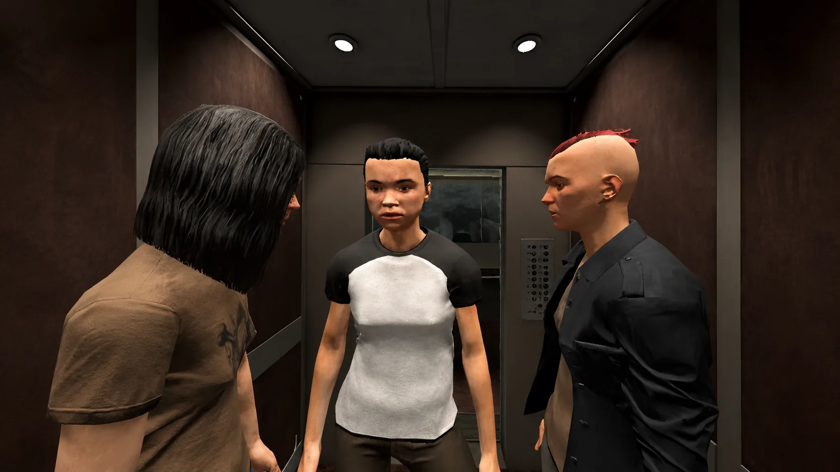
- Standard lift with other occupants (55-60). Reduces the volume available per person and adds the inability to move freely.
- Standard lift with a brief stop between floors (60-70). The hinge level of the protocol: this is where the core cognition of being trapped shows up.
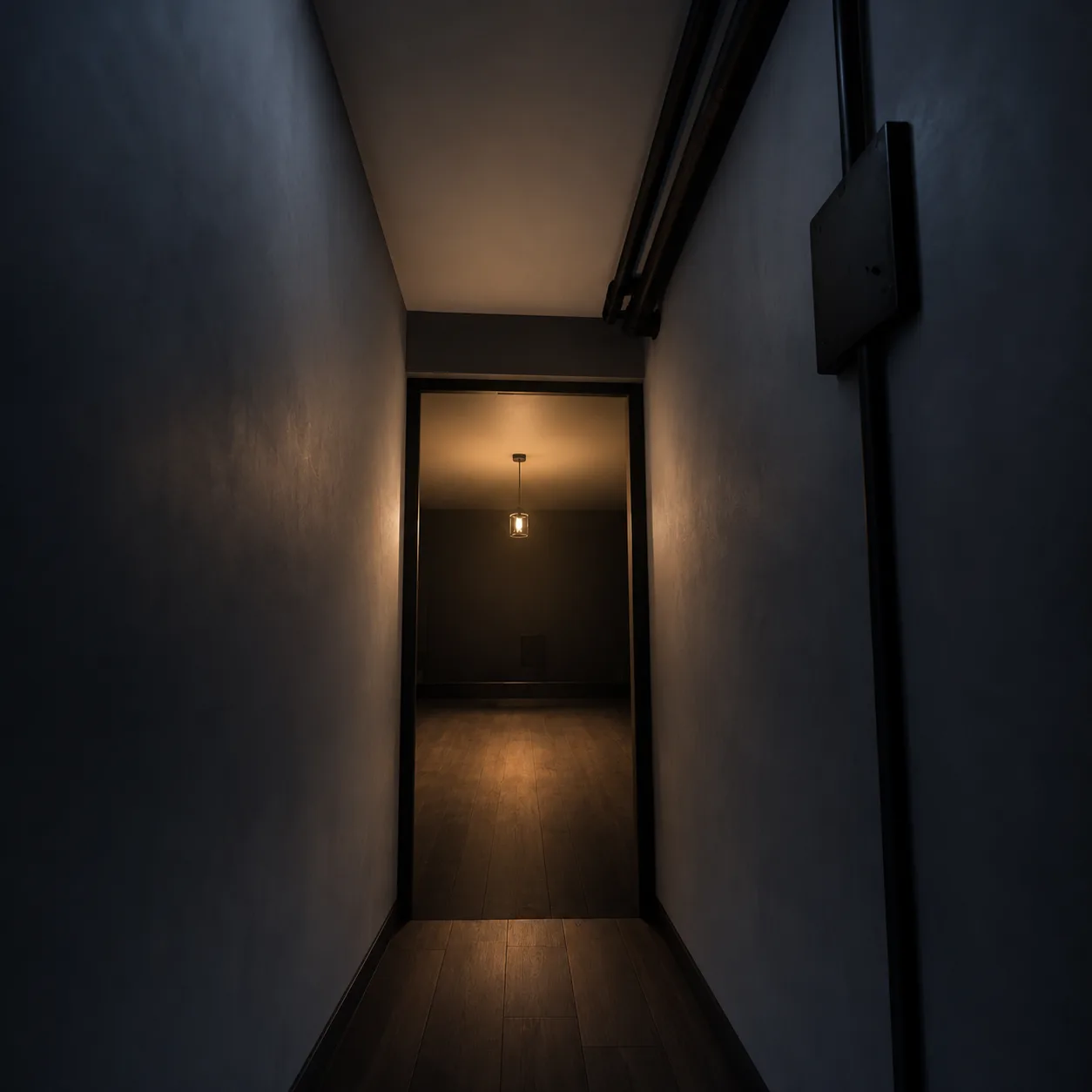
- Narrow tunnel or corridor on foot, exit visible (65-75). Changes the type of enclosure and forces the learning to generalise beyond the lift.
- Narrow tunnel, exit not visible, reduced lighting (75-85). Both axes at maximum simultaneously.
- Scanner-type enclosure: supine, very tight space, intermittent noise, duration not announced (85-95). The level built for the patient referred from radiology.
Two notes on use. First: you do not move up a level because a session finished, but on a habituation criterion within the level, with two consecutive trials in which peak subjective distress falls clearly relative to onset. Second: levels 6 and 8 carry most of the useful therapeutic work, and deserve whole sessions rather than being passed through on the way somewhere else.
In the VRET claustrophobia scenario, levels 3 to 6 run on the lift cabin, with ceiling height, width and stop duration as independent controls from the clinician panel, and levels 7 and 8 run on the tunnel.
The patient referred from radiology: claustrophobia and MRI
There is a referral profile that arrives with an unusual feature: a deadline. The patient has an MRI scan booked, has abandoned one or two previous appointments, and the radiology department has suggested they seek help before trying again.
This case forces a rethink of the therapeutic goal. The task is not to resolve the claustrophobia, but to produce enough tolerance for a single, scheduled exposure of known duration. That is a more modest and entirely legitimate objective, and it is worth stating explicitly with the patient in the first session so they do not read discharge as an unfinished treatment.
The format that fits best is short and tightly directed:
- Session 1. Assessment, formulation and psychoeducation on the anxiety response. Visor acclimatisation and a level 1 trial.
- Session 2. Levels 3 to 6, with emphasis on work under unknown duration.
- Session 3. Level 9 replicating the real conditions of the scan: supine, arms at the sides, intermittent noise and an instruction to stay still.
- Session 4. Level 9 repeated, rehearsing the coping plan agreed with the department: an agreed signal, paced breathing, attentional focus.
Two things are worth flagging to the referring team. One, the instruction to stay still is a stimulus in its own right and usually scores higher than the bore diameter itself; if it is not rehearsed, the preparation is incomplete. Two, the real duration of the scan should be known before session 3, because the rehearsal loses value if the patient calibrates for twenty minutes and the examination runs to forty.
This profile is also a stable and under-exploited referral route. If you are weighing up building a service line around one specific presentation, the article on how to build a profitable phobia-specialty VR practice works through the commercial reasoning in full.
Errors that flatten the habituation curve
Four mistakes account for most claustrophobia hierarchies that stop progressing.
Withdrawing the patient at the peak. Ending exposure while subjective distress is at its maximum teaches the opposite of what was intended. The exit criterion is the decline, not the clock and not the therapist's discomfort.
Missing subtle safety behaviours. In claustrophobia they are discreet and easy to overlook: staring at the door, counting silently, keeping a finger on the button, controlling the breath deliberately. All of them block disconfirmation of the expectancy. It is worth asking about them explicitly at the close of every trial, because patients rarely report them spontaneously.
Trials that are too short. A three-minute trial at level 6 gives the curve no time to come down. Time inside is the active ingredient, and fewer levels with longer stays beat a quick run through all nine.
Mistaking visor discomfort for anxiety. A patient uncomfortable from weight, heat or optical misalignment will score high without that reflecting phobic activation. Ruling it out at level 1 is quick and avoids misreading the whole hierarchy that follows. The guide to VR motion sickness, its causes and prevention covers the pre-session fit, and the visor hygiene protocol between patients covers the turnaround.
When to move to in vivo exposure
Virtual exposure does not stand in for in vivo work: it makes it possible. The aim is to reach the real lift with the patient having already disconfirmed the core expectancy, rather than reaching it never having been there at all.
Three reasonable criteria for scheduling the move: the patient completes level 6 with a clear decline across two consecutive trials; they have identified and dropped their main safety behaviours; and they can articulate what they expected to happen and what actually happened. That third criterion is the best predictor of generalisation, and it is qualitative.
On record-keeping, three data points per trial are more than enough to support the clinical note: subjective distress at onset, peak and close; time spent inside; safety behaviours observed or reported. The VR practice setup checklist sets out the minimum record per session.
On treatment-length expectations, the article on how many VRET sessions each presentation realistically needs gives ranges by condition that are worth sharing with the patient in the first session.
This article is for informational purposes for psychology professionals. It is not clinical advice for any individual case and does not replace the judgment of the licensed psychologist in charge. VRET is professional clinical-support software, not a CE-marked medical device.
Frequently asked questions
How does claustrophobia differ from agoraphobia?
In the core fear, not in the setting. In claustrophobia the patient fears being trapped or running out of air, and the focus is on the physical properties of the enclosure. In agoraphobia they fear incapacitating symptoms with no escape and no help available, and the focus is on the accessibility of rescue. A useful clinical contrast: if anticipatory anxiety drops once the patient knows they will have company and that the door opens in five minutes, the agoraphobic component dominates.
Is claustrophobia only a fear of enclosed spaces?
No. Physical confinement is only one of the two axes. Perceived control, meaning whether the patient knows how long it will last, whether they can get out on their own and whether anyone is outside, moves subjective distress as much as or more than the volume of the enclosure. Two situations with identical tightness can differ by thirty points depending on what the patient knows about duration.
Can virtual reality be used to prepare a claustrophobic patient for an MRI scan?
It is one of the referral profiles where brief preparation fits best, because the objective is bounded: tolerating a single, scheduled examination of known duration. A four-session format ending with rehearsal in the supine position, with intermittent noise and an instruction to stay still, reproduces the critical conditions. It is worth agreeing the objective with the patient and confirming the real duration of the scan with the referring department.
How many sessions does a patient with claustrophobia need?
It depends on the objective. Bounded preparation for one specific medical procedure usually resolves in three or four sessions. Treating the full presentation, generalising to lifts, tunnels and transport, runs longer and depends above all on how much time the patient spends at levels 6 and 8 of the hierarchy. Short trials lengthen treatment more than initial severity does.
What should I do if the patient removes the visor mid-exposure?
Record it as clinical data, not as failure. It indicates the level sat above the working window, or that an undetected safety behaviour appeared. The usual response is to step back one level, extend time spent there until you obtain a clear decline across two trials, and review what the patient expected to happen at the moment they pulled out.
Keep reading
Acrophobia vs Vertigo: A Clinical Differential Guide
Acrophobia, vestibular vertigo and visual height intolerance get confused in practice. Five intake questions, two questionnaires and clear referral criteria.
Specific phobiasAmaxophobia: VR Exposure Therapy for Driving Phobia
How VR exposure treats driving phobia when in-vivo practice is logistically complex: a graduated protocol, clinical evidence, and session structure for licensed psychologists.
Specific phobiasSocial Anxiety Treatment: Why VR Succeeds Where Imagery Fails
Clinical limits of imaginal exposure in social anxiety and what VR adds: graded avatars, eye contact, hostility levels, and trial evidence (Anderson, Bouchard, Kampmann).
VRET is professional clinical-support software, not a CE-marked medical device. Clinical supervision remains with the licensed psychologist in charge.